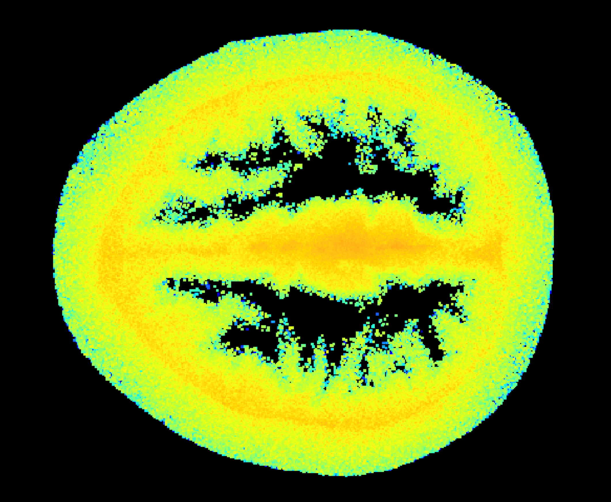
Imaging through the brain
Portable detection of head injuries, strokes and brain disorders
Researchers at the University of Glasgow are developing imaging techniques which have the potential to create cheap, portable imaging cameras to see through the human body, and in particular, the brain.
Traditional methods for imaging inside the body or the brain require expensive and invasive techniques such as fMRI or CT-xray scans.
Using new portable imaging cameras could allow GPs and paramedics to diagnose fractures, head injuries, and strokes on the spot or carry out basic autism and Alzheimer checks.
The research uses a light-in-flight technique to detect photons (light particles) transmitted through the human head. Researchers are combining single-photon detection with precise time resolution and machine learning techniques to image through highly diffusive media such as tissue.
Results so far show that images can be recovered through many 10’s mm of highly-scattering, tissue-like material obtaining mm resolution of buried objects. The goal is to achieve MRI-like image quality using tissue simulations showing that it might be possible to non-invasively image the outer 10s mm of the brain.
The team are also developing AI techniques to further improve fMRI capability.

Benefits
- Low-cost, portable alternative to MRI or CT-Xray
- Early, on-site diagnosis of stroke (in GP surgery or ambulance)
- Less invasive compared to traditional scanning techniques.
Applications
- Primary healthcare settings such as GP surgeries
- Secondary healthcare settings such as hospitals as an alternative to more expensive and invasive

Healthcare
Meet our investigator
Professor Daniele Faccio is a Royal Academy Chair in Emerging Technologies, Fellow of the Royal Society of Edinburgh and Cavaliere dell'Ordine della Stella d'Italia (Knight of the Order of the Star of Italy). He joined the University of Glasgow in 2017 as Professor in Quantum Technologies where he leads the Extreme-Light group.

